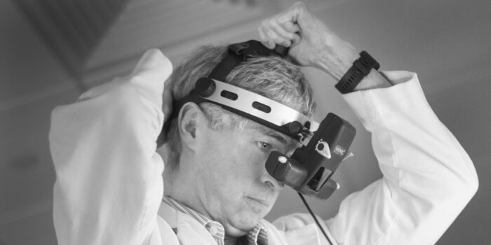
As leaders in retina care we use the most advanced diagnostic and imaging equipment and software available. Click through to find information on our diagnostics.
Fundus autofluorescence is a non-invasive diagnostic test that involves taking digital photographs of the back of the eye without a contrast dye. These images can help with the early detection of diseased retina in serious eye conditions such as macular degeneration, hereditary retinal degenerations, and retinal toxicity from the long-term use of medications such as hydroxychloroquine (plaquenil).
An electroretinogram (ERG) is a painless diagnostic procedure that evaluates the function of the retina, the light-sensitive lining on the back of the eye where light is focused. This test can aid in the diagnosis of several different retina conditions that can lead to serious complications, including permanent vision loss.
Fluorescein angiography is a classic diagnostic test that involves taking photographs of the blood vessels in the eye with the help of a contrast dye. Fluorescein is a yellow dye that allows the retina to glow when exposed to a certain wavelength of visible light.
Indocyanine green angiography is a diagnostic test that involves taking photographs of the blood vessels in the eye with the help of a contrast dye. Indocyanine is a green dye that works with infrared light and is visualized with a special digital camera.
Microperimetry is a type of visual field test which uses one of several technologies to create a “retinal sensitivity map” of the quantity of light perceived in specific parts of the retina. Visual field testing is widely used to monitor pathologies affecting the periphery of vision such as glaucoma.
Optical coherence tomography (OCT) is a non-invasive way for your physician to visualize the microscopic structures of your retina. The technology has revolutionized our care for patients with conditions affecting the retina, such as macular degeneration, diabetic retinopathy, retinal vein occlusions, and many more.
Optical coherence tomography angiography (OCTA) is a non-invasive way for your physician to visualize the vascular structures of your retina. With OCT-A, high definition digital images of the blood vessels can be obtained in seconds and analyzed immediately thereafter.
Ocular ultrasonography (“ultrasound”) is a non-invasive diagnostic imaging test that utilizes the movement of sound waves to generate an image of internal structures within the eye. It uses the same technology as an obstetrical ultrasound that is used to examine a baby in the womb, but in a smaller format designed for the eye.